
Thanks to the contribution of the Fondazione Roma, through the Biomedical University Foundation, as part of a broader funding for the development of the Center for Research and Treatment of Alzheimer’s Disease at FPUCBM and UCBM.
On March 27, 2024, the inauguration ceremony of the new 3 Tesla Magnetic Resonance Imaging (MRI) machine of the Fondazione Policlinico Universitario Campus Bio-Medico took place. This new equipment will allow for greater spatial resolution, providing increasingly detailed images, and, in some cases, faster examination compared to the more common 1.5 Tesla MRI machines. The higher resolution of the instrument enables the identification of smaller lesions and anatomical structures that may not be visible with machines from previous generations, particularly in the diagnosis of brain or musculoskeletal pathologies and spinal conditions. The quality of the images makes this equipment the reference for examinations of cerebrovascular and neurological districts, as well as enabling functional, spectroscopy, prostate, and pancreatic examinations.
The acquisition of the equipment was made possible thanks to the contribution from Fondazione Roma, through the Biomedical University Foundation, as part of a broader funding for the development of the Center for Research and Treatment of Alzheimer’s Disease of the Fondazione Policlinico Universitario Campus Bio-Medico and the University Campus Bio-Medico of Rome.
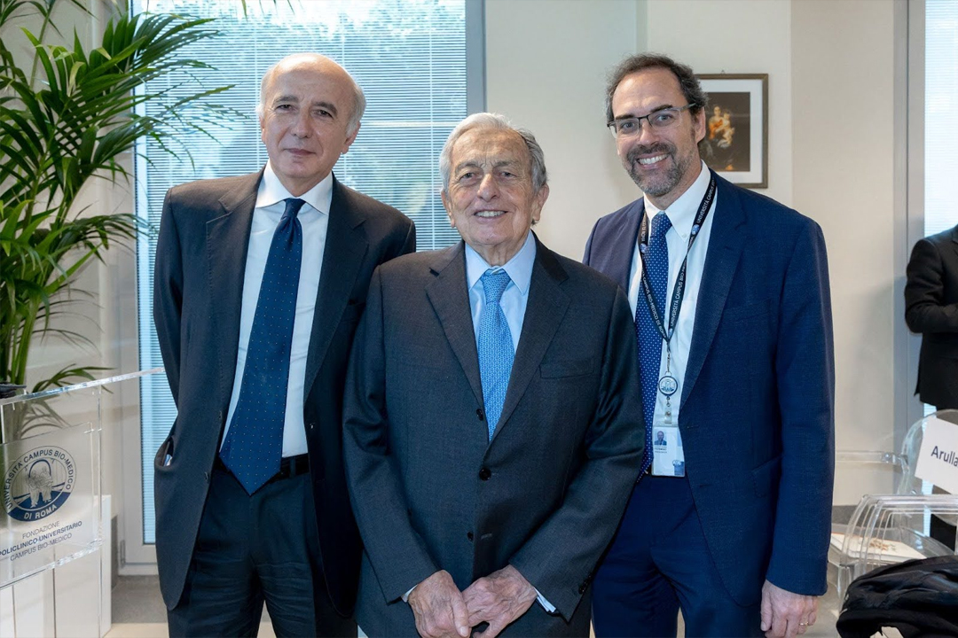
In addition to the leaders of the Fondazione Policlinico Universitario Campus Bio-Medico, the inauguration was attended by Prof. Bruno Beomonte Zobel, coordinator of the Imaging Center of the Policlinico Universitario Campus Bio-Medico, Franco Parasassi, President of Fondazione Roma, Prof. Paolo Arullani, President of the Advisory Board of the Biomedical University Foundation, Alessandro Pernigo, President of the Biomedical University Foundation, Lawyer Civita Di Russo, Deputy Chief of Staff of the President of the Lazio Region, and Hon. Ylenja Lucaselli, chair of the Budget, Treasury, and Programming Committee of the Chamber of Deputies.
Eng. Carlo Tosti, President of the Fondazione Policlinico Universitario Campus Bio-Medico, stated: “Today is an important opportunity to network with the Regional Health Service, considering that this new equipment will be accessible to citizens based on regional planning and will cover various clinical areas. It represents a significant investment that reflects, thanks to its excellent characteristics, the motto of the Fondazione Policlinico Universitario Campus Bio-Medico ‘Science for Humanity,’ guaranteeing high levels of care and assistance while always considering the individual in all their needs and dimensions.”
Franco Parasassi, President of Fondazione Roma, stated: “With today’s inauguration, we begin to give concrete substance to the path that will lead, in the coming years, to the completion of the Integrated Research and Treatment Center for Alzheimer’s Disease – Fondazione Roma presented last September and to which all the involved parties are working diligently, with enthusiasm and full awareness of the innovative contribution it will make to early diagnosis and research, appropriately combined here, of this neurodegenerative pathology to combat which Fondazione Roma has been engaged, on various intervention levels, for many years.”
MRI machines with three Tesla magnets, compared to those with 1.5 Tesla, provide superior performance, particularly in the study of the pituitary gland, hippocampus, brainstem nuclei, cranial nerves, inner ear, cerebral cortex, and tumors. Additionally, they reduce the risk of distorted images, thus eliminating the need for additional MRIs.
Prof. Bruno Beomonte Zobel, Coordinator of the Imaging Center of the Fondazione Policlinico Universitario Campus Bio-Medico, explained: “With this type of equipment, advanced techniques such as MRI Spectroscopy, Diffusion Tensor Imaging, MRI Tractography, and Functional MRI with BOLD signal can be used. Functional MRI can identify areas of the brain that consume oxygen while the subject being examined with the MRI machine is performing a certain activity such as watching a video, listening to music, or making a small movement with a finger. In this way, it has been possible to study, in healthy volunteers, which brain areas are involved, in the sense that they are activated and consume oxygen, in making certain decisions.”